Science of Microspectroscopy
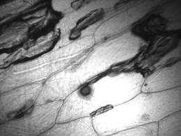
Ultraviolet image of an onion cell
Microspectrophotometer Operation
Uses of Microspectrophotometers
Algorithms of Microspectroscopy
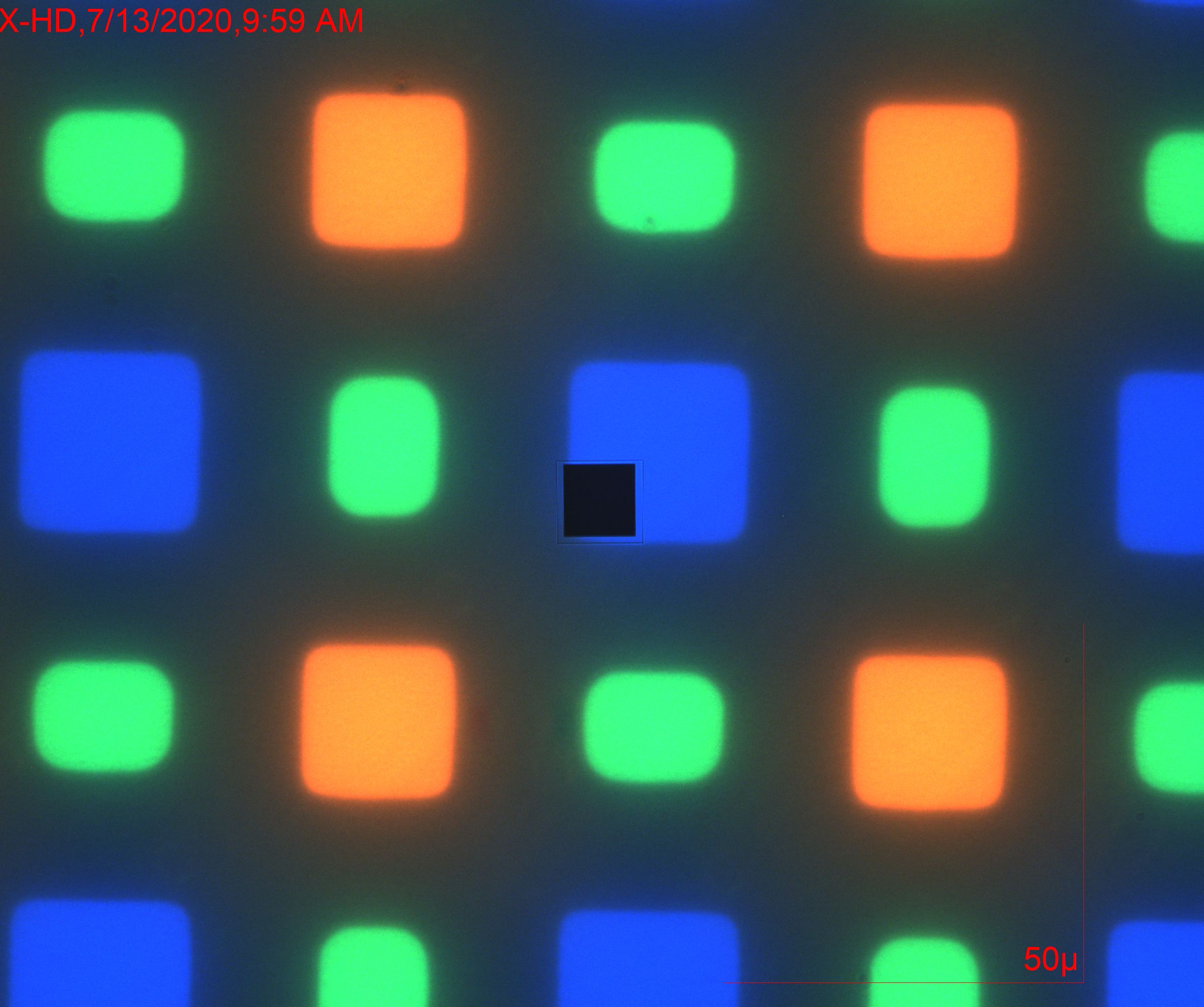
2030PV PRO™ Microspectrophotometer
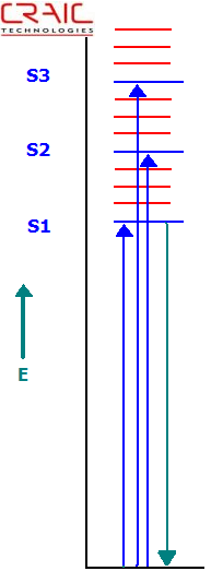
Jablonski Energy Level Diagram Depicting Absorbance and Fluorescence Transitions
The UV-visible-NIR microspectrophotometer is designed to measure spectra of microscopic areas or microscopic samples. It can be configured to measure the transmittance, absorbance, reflectance, polarization and fluorescence of sample areas as small as one micron.
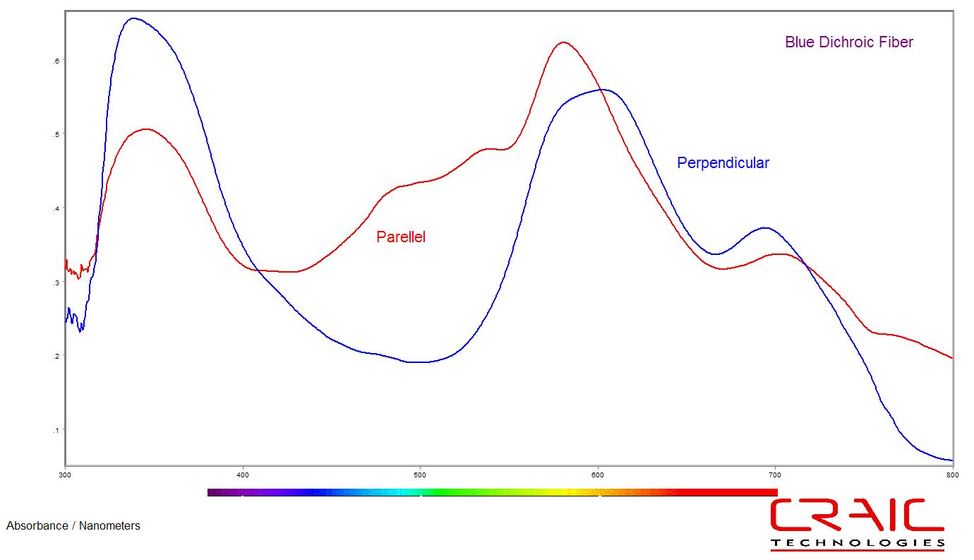
Absorbance
Microspectroscopy is the study of the quantized energy transfer between radiation and matter. The energy of light is directly related to its wavelength by the Einstein-Planck relationship:
The electromagnetic energy of a photon is inversely proportional to its wavelength. In other words, short wavelength or blue light is higher energy than red light. Due to the differences, the light causes different effects when it interacts with molecules. In the ultraviolet-visible region, electronic transitions are mainly observed. Namely, when a photon of the proper energy is absorbed by a molecule, an electron is excited to higher energy level or shell. This is most commonly described in what is called a Jablonski diagram (as shown to the left).
For a photon to be absorbed, the energy of the photon must correspond to the difference in energy between the ground state and the excited state to which the electron transfers. As can be seen in the diagram, the electron jumps to the first excited state (S1) when an electron of the corresponding energy is absorbed. If a photon of a higher energy, one that corresponds to the difference between the ground and say the second excited state (S2), were absorbed, the electron would jump the S2 state.
This process is called absorbance.
The energy levels of the molecules are due to the types of atoms and how they are bound to one another. Additionally, the shape of the molecule as well as its environment can also play a part in structuring the energy levels. In fact, a dye chemist can "tune" a dye molecule by adding or removing functional groups or atoms of a molecule, thereby changing its color.
FluorescenceAfter the electron has jumped to the excited state, it then decays by internal conversion to the lowest excited state, S1. From there it can decay back to the ground state by a number of paths. The most common is a radiation-less transition whereby the electron drops from the S1 excited state to the ground state losing energy without the emission of a photon. However, when the electron drops from the S1 state to the ground state with the emission of a photon, the process is called fluorescence. It is a rapid process and fluorescence lifetimes usually follow first order kinetic rules. It should be noted that the fluorescence intensity is governed by many factors, some of which include excitation wavelength, quantum yield, quenching materials and even molecular structure.
Reflectance
Reflectance microspectroscopy simply measures the spectra of electromagnetic energy reflected from the sample. The portion that is not reflected may have been absorbed (see above) or transmitted through the sample (if transparent to that wavelength of light) or even scattered. Reflected light may be divided into two types: specular and diffuse. Simply put, specular reflectance is like the reflectance from a mirror. The light is reflected at the same angle as it impinges upon the mirror surface. Diffuse reflectance is similar to what occurs with white paper. Light is effectively reflected at all angles.
Chemistry and ColorSubstances absorb, reflect or emit light in ways that are dependant upon their chemical structure and their environment. Examples include the pigments found in automotive paints that reflect specific wavelengths of light which we perceive as a color...or quantum dots, commonly used for biological analysis, that emit light of specific wavelengths when excited and then decay back to the ground state with the emission of photons. The optical properties of these colorants depend upon the molecular structure of their chromophores (the molecules that interact with the light) as well as their environment. Measuring the optical properties of these molecules on the microscale is called microspectroscopy.